To produce a three-dimensional image of a patient, medical professionals can use a technique of scanning utilizing positron emission tomography principles. In its most basic sense, this system is used to detect gamma rays introduced into the body at a molecular level.
Basic Function of Positron Emission Tomography
At its base, positron emission tomography principles use a variety of techniques developed using nuclear technology. A patient is injected with radioactive isotopes via the circulatory system. These isotopes are most commonly short-lived flurodeoxyglucose, a type of sugar, and will break down over a brief period of time. After injection, the patient needs to wait for as the tracer isotopes enter the tissues of interest and become concentrated elements within the molecules of the region. This usually takes about an hour.
As the radioactive elements decay, they undergo positron emission known as beta decay. Essentially, the antiparticle of the electron, positrons, are emitted, eventually coming into contact with each other and becoming annihilated. This produces gamma photons that move in different directions. The PET scan technique is able to identify these gamma rays and make an image of the molecules.
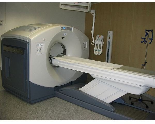
Above right: PET and CT Scanner. (Supplied hg6996 at Wikimedia Commons; Public Domain; https://upload.wikimedia.org/wikipedia/commons/c/c4/16slicePETCT.jpg)
How Gamma Rays Are Detected
The scanning device uses a material known as a scintillator to detect the gamma rays produced through the positron emission tomography principles. The scintillator becomes luminescent when the gamma photons are detected. This is then isolated by devices which detect a variety of light along the electromagnetic spectrum known as photomultiplier tubes. A computerized system then processes the location and image of each gamma ray, giving a picture of the subject’s section of interest. Because the entire system relies on the photons traveling in opposite directions after the mutual annihilation of the positron and electron, only signals resulting from this feature of motion are detected. Any gamma photon that does not travel in an exact opposite pattern of another is treated as an anomaly and ignored.
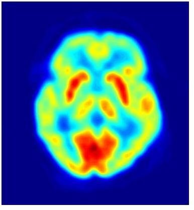
Above left: PET image. (Supplied by Jens Langner at Wikimedia Commons; Public Domain; https://upload.wikimedia.org/wikipedia/commons/c/c6/PET-image.jpg)
Overall Challenges of Positron Scanning
One of the major challenges with positron emission tomography principles is the technique of reconstructing the image of the patient. Since the technology uses concepts such as the vacuum tube used to identify the luminescence seen by the scintillator, developing a three or four-dimensional representation can be difficult. Many researchers use this technology in conjunction with other forms of body scanning , such as CT scans. Some machines have even been constructed that use both concepts within the same procedure, allowing patients and physicians to accomplish two goals in the same amount of time.
Development of Positron Emission Tomography Principles
During the 1950s, David E. Kuhl and Roy Edwards worked at the University of Pennsylvania in an effort to use the positron and electron annihilation features as an identification process. This research continued at a number of different facilities throughout the next few decades, ultimately leading to the synthesis of the radioactive isotope carriers needed for proper identification within the human body. In August 1976, the first patients were tested using positron emission tomography principles at the University of Pennsylvania. Ultimately, this was adapted to the modern methodology, giving scientists a new way to scan the body.
Resources
“Nuclear Medicine” Radiology: https://www.radiologyinfo.org/en/info.cfm?pg=PET
“Positron Emission Tomography” WebMD: https://www.webmd.com/a-to-z-guides/positron-emission-tomography


