The Golgi apparatus is a part of the cellular structure that assists in the modification and delivery of proteins and other macromolecules. Made up of flat membranes called cisternae, the Golgi apparatus also protects against cell destruction known as apoptosis.
Found in most eukaryotic cells , the Golgi apparatus – also known as the Golgi Body – was first discovered by Camillio Golgi. Golgi was an Italian physician who was able to identify the organelle in 1898 using a staining technique that fixed silver chromate particles to membranes.
Golgi Apparatus Function
The main function of the Golgi apparatus is to be responsible for handling the macromolecules that are required for proper cell functioning. It processes and packages these macromolecules for use within the cell or for secretion. Primarily, the Golgi apparatus modifies proteins that it receives from the rough endoplasmic reticulum, however, it also transports lipids to vital parts of the cell and creates lysosomes. As part of eukaryotic cells, the Golgi apparatus works in unison with the endomembrane system.
Other functions of the Golgi apparatus include the production of glycosaminoglycans, which go on to form parts of connective tissues. The Golgi will use a xylose link to polymerize the glycosaminoglycans onto proteins to form proteoglycan.
It then performs sulfation onto the proteoglycans in order to aid in signaling abilities and giving the molecule a negative charge.
The Bcl-2 genes that are located within the Golgi apparatus also play a significant role in preventing apoptosis, or the destruction of the cell.
Golgi Apparatus Structure
The Golgi apparatus has a structure that is made up of cisternae, which are flattened stacks of membrane usually found in a series of five to eight. These cisternae help proteins travel from different points in the cell using enzymes . In order to modify a macromolecule, cisternae’s enzymes need the addition of carbohydrates and phosphates to properly label each protein for its ultimate destination. These carbohydrates and phosphates are received by the Golgi apparatus through nucleotide sugars delivered to the organelle from the cytosol.
How the proteins and vesicles pass through the Golgi apparatus structure is unclear, however, there are theories regarding the subject. According to the vesicular transport model, there are a variety of compartments located between the cis, essentially the beginning of the Golgi apparatus, and the trans, the end. These compartments shuttle along the macromolecules from section to section using membrane-bound carriers. The cisternal maturation model states that the vesicles fuse to each other at the cis face of the Golgi apparatus and are essentially pushed along as new vesicles fuse together behind them.
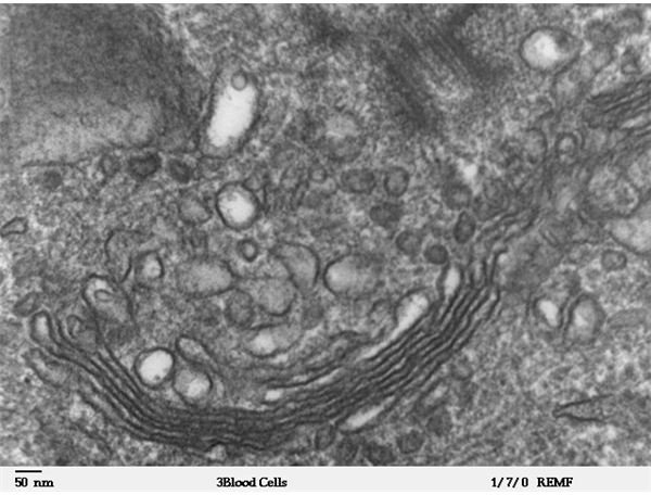
Above: A high magnification transmission electron microscope image of a human leukocyte, showing golgi (Image credit: Louisa Howard at Wikimedia Commons, https://en.wikipedia.org/wiki/File:Human _leukocyte,_showing_golgi_-_TEM.jpg_constellation_map.png, GNU Free Documentation license.)


