A compound microscope is basically a device capable of magnifying microscopic elements that may be invisible to normal human eyes. Let’s understand how to adjust and use a compound microscope.
A microscope can be considered one of the greatest inventions and contributions to medical science. A microscope is an optical instrument with sets of intricately devised lenses capable of magnifying anything that may be impossible to visualize through normal human eyes.
Specifically these devices are utilized in laboratories to examine various harmful parasites or microbes like bacteria and viruses, as well as other biological specimens, including all types of human tissue.
Just like any other instrument a microscope too may have its own limitation as far as magnifying is concerned and is termed as the resolving power or the resolution of a microscope. An ordinary microscope normally may be quite inefficient in providing comprehensive details regarding a microbe or an infected tissue due to its low resolution. To overcome this a compound microscope is usually preferred which may have a resolution or a magnifying capacity of more than 1000 times. A small in-built light source is normally incorporated inside them for illuminating the specimen under scan and hence they are also known as compound light microscopes.
Their resolving power is typically around 0.2 µm, the wavelength of visible light being just about the double of this figure. Images obtained from this type of microscope are called photomicrographs.
The following section discusses how to adjust and use a compound microscope.
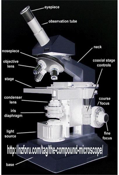
Setting Up the Microscope
The following stages are normally employed to set up a compound light microscope:
-
Initially, the resolving nose piece is moved to suitably lock the lower objective lens into position.
-
Next, looking at the objective lens and the stage sideways, the coarse focus shaft is turned to make the stage move near the objective and as close as possible to the slide without making physical contact.
Advertisement -
The light source and the diaphragm are now adjusted until optimum illumination is reached.
-
After this the coarse focus is gently turned to move the stage downwards until the view shifts into a broader focus. The fine adjustment control is further optimized for a perfect focus.
Advertisement -
Finally, the specimen slide is moved until it appears at the center of the frame, again the illumination and the diaphragm is adjusted to improve clarity.
How a Compound Microscope is Used in Laboratories
The following couple of test methods (normally employed for the diagnosis of disease causing pathogens) and the acquired test results using a compound microscope simply proves its efficiency and indispensability in the medical field. The examples will also help you to understand how to prepare a piece of tissue of examination under a compound light microscope.
Let’s analyze the utility of a Compound Light Microscope (CLM) through the following illustrations:
Identifying a Yeast Colony
**

The microscopic slide is first sanitized using sterile water,
A very small quantity of the specimen colony is diluted in a drop of sterile water using a sterile bacteriological inoculating loop,
The prepared slide is covered with a glass coverslip,
The slide is then inserted under a CLM for examination. It will be important to note that for a wet mount specimen an oil immersion lens is never used.
Observation: The magnified image of the sample slide under the CLM normally yields the following facts:
Yeast cells are more easily distinguishable due to the major difference in their size and shape from the bacterial cells.
They are much larger - about 10 times more than the bacterial cells, moreover yeast cells may be found to be oval in shape unlike the bacterial cells which are mostly elongated.
The important identifiable feature among yeast cells are their budding phases which is never found in bacterial cells.
You may also find that the heat of the CLM light source causes the yeast cells to wiggle. This “wiggling” of the cells is technically termed as the Brownian motion. However this may happen only if the examinations are done for prolonged periods.
Diagnosing Vaginal Infections
**
A sample of the vaginal discharge is acquired over a sterile swab,
The specimen material in the swab is gently brought in contact with the sterile saline and stirred to let them mix,
Almost immediately the preparation is covered with the glass coverslip so that it DOES NOT dry up,
The above wet mount is investigated under a CLM in a rather reduced illumination (Do not use oil immersion lens for wet mounts),
Observation: The CM magnifications may normally reveal the following possible causes of infection:
Presence of yeast cells, indicating colonies of yeast vaganities,
If motile trophozoites of T. vaginalies are seen, the presence of trichomoniasis may be presumed,
If an accumulation of clue cells (squamous epithelial cells occupied with colonies of cocobacilli) is witnessed, will indicate a bacterial vaginosis.
In the above case a Gram stain preparation of the vaginal fluid is recommended and its inspection under the CM may specify the following results:
Presence of some Gram-positive bacilli or Lactobacillus spp.
Presence of many Gram-negative bacilli or Bacteroids spp. Many curved Gram-negative bacilli or Mobiluncus spp.
Presence of Gram-variable coccobacilli or Gardnerella vaginalis.
Identification of Anthrax Bacteria

References
https://books.google.co.in/books/


