Has your doctor scheduled an echocardiogram for you or a loved one? If so, read on to learn more about this diagnostic test.
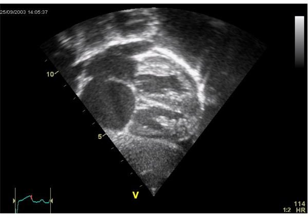
An echocardiogram, also sometimes called an “echo”, is a diagnostic test performed to provide a graphic outline of how the heart is moving. It is a type of ultrasound, using high-frequency sound waves, to create pictures of the heart’s chambers and valves. This gives the sonographer (the technician performing the test) the ability to get a good look at how well the heart is pumping. This test is often done along with a color Doppler ultrasound and Doppler ultrasound to see how well the heart’s blood is flowing across the valves of the heart.
Why is an Echocardiogram Performed?
This diagnostic test is performed for a variety of reasons. The most common reasons include:
- Determining how well the heart is functioning overall
- To look for several different types of heart disease
- To determine how effective surgical or medical treatments were
- To keep an eye on how heart valve disease is progressing over time
What Does a Patient Need to do to Prepare for this Test?
There is no preparation for an echocardiogram for almost all patients. Patients should eat, drink, and take all of their medications as they would on any other day. If for some reason the patient’s doctor feels the patient should prepare in any way they will let them know in advance.
What to Expect During an Echocardiogram
Patients will most often change into a hospital gown and take off all of their clothing above the waste. They should also remove all jewelry. The cardiac sonographer (the technician performing the test) will place three electrodes on the patient’s chest and then these electrodes will be attached to a machine known as an electrocardiograph monitor (EKG or ECG). This machine will track the electrical activity of the patient’s heart. The sonographer will have the patient life in their left side and use a sound-wave transducer (similar to a wand) on several different places on the patient’s chest using a little bit of gel. The patient may need to reposition several different times throughout the echocardiogram so that the sonographer can get all of the pictures that they need to get. The patient may also need to briefly hold their breath during certain times throughout this test to help the sonographer get certain pictures.
An echocardiogram will take about 40 minutes, on average, to complete. Patients should not experience much discomfort except some light pressure when the sonographer is using the transducer and some coolness when the gel they use touches the skin.
Once the test is over the patient can get dressed, go home, and resume their normal activities.
Resources
Medicine Net. (2007). Diagnosing Heart Disease: Echocardiogram. Retrieved on April 29, 2010 from Medicine Net: https://www.medicinenet.com/echocardiogram/article.htm
Medline Plus. (2009). Echocardiogram. Retrieved on April 29, 2010 from Medline Plus: https://www.nlm.nih.gov/medlineplus/ency/article/003869.htm
Image Credits
Echocardiogram: Ekko – Wikimedia Commons


