The nerve cell structure of neurons are highly specialized and designed to specifically receive, process and relay electrochemical signals from one cell to another. When these cells are placed in a series, each part is perfectly positioned to function in unison for the nerve system.
Soma
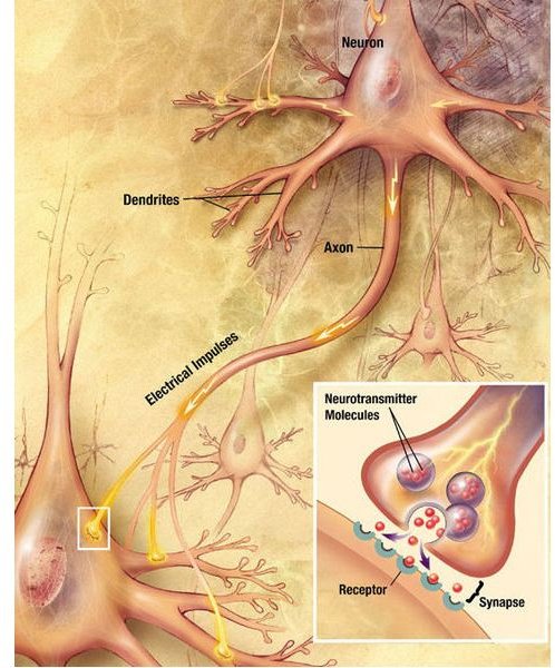
The nerve cell structure is based around the soma, the part of the cell which contains the nucleus. The soma acts as the basic body of the nerve cell structure. All secondary parts of the neuron are based around the soma, although unlike other cell types, the nucleus is not located at the center of the cell. The RNA of the nerve cell is located within the nucleus. The messenger RNA exists around the nucleus, producing proteins that make the cells and nervous system in general function. Soma range in size from four micrometers all the way up to a single millimeter in larger invertebrates.
Above right: Nerve cell. (Supplied by the US National Institutes of Health; Public Domain; https://upload.wikimedia.org/wikipedia/commons/3/30/Chemical _synapse_schema_cropped.jpg)
Dendrites
Extending from the soma are branches of biological material known as dendrites. These branches appear similar to the limbs of a tree. The main purpose of the dendrites in the nerve cell structure is to receive electrochemical stimulation from other neurons. Stimulation from one cell to another travel across the synapses, areas between each cell. The electrical stimulation makes contact with the dendrites and the signal is sent to the soma for processing.
Right: Neuron. (Supplied by Medlat at Wikimedia Commons; Creative Commons; https://upload.wikimedia.org/wikipedia/en/3/32/Smi32neuron.jpg)
Axon

The soma also is connected to another projection known as the axon. This resembles a long cable that extends in diameter much further than the soma itself. This distance can be anywhere from ten times the length of the soma, all the way to tens of thousands. Between the axon itself and the soma is a portion of the neuron known as the axon hillock, the most sensitive portion of the nerve cell due to the high volume of sodium channels.
The axon itself is divided into three specific components. Covering the entire axon is a material known as the myelin sheath, an insulating layer which prevents the electrical signals from being lost as they flow through the axon. Essentially, the myelin sheath acts as the rubber that covers and electrical chord. The actual conduction of the signal is handled by a part of the axon known as the Schwann cell. In addition to sending signals, these are also responsible for regeneration and overall development of the nerve. In between each Schwann cell is a gap connecting the axon together, known as nodes of Ranvier. These nodes are not insulated with the myelin sheath, meaning additional electrical signals can be generated at these points.
Above right: Cultured Schwann cell. (Supplied Ucbtbej at Wikimedia Commons; Creative Commons; https://upload.wikimedia.org/wikipedia/commons/7/75/Cultured _schwann_cell.jpg)
Axon Terminal
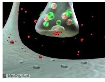
The final component of the nerve cell structure is the axon terminal. These are similar to the dendrites, however, they have the opposite purpose. The axon terminal receives the signal from the rest of the neuron and sends it along the long branches across the synapse and to the dendrites of another cell. This is how nerve cells connect to each other and communicate as the overall nervous system.
Left: Axon Terminal. (Supplied by Cushlash at Wikimedia Commons; GNU Free Documentation License; https://upload.wikimedia.org/wikipedia/en/c/cd/BioDigital _Exocytosis.jpg)
References
- “The Nerve Cell in General” Fortune City: http://www.fortunecity.com/greenfield/buzzard/387/nervecellgen.htm
- “Nerve Cells” Hyperphysics: http://hyperphysics.phy-astr.gsu.edu/HBASE/Biology/nervecell.html


