The mechanisms and workings of MRI machines are based on the principles of nuclear technology. Our basic understanding of how subatomic particles interact with magnetism and radio frequencies allows us to utilize the design of the MRI machine to conduct detailed scans of biological tissue.
Basic Functions of MRI Machines
Since the body is composed largely of water, a magnetic resonance scan can isolate the molecules from other parts of the body to create an image. The mechanisms and workings of MRI machines rely on a large magnet to produce a electromagnetic field focused on the subject’s area of interest. The nuclei and protons of the water molecules then become aligned with the direction of the field. A radio frequency field is then projected for a brief period of time onto the same area, causing the protons to shift alignment. The scanner is able to pick up these changes, giving a technician the information necessary to determine the location of the molecules.
Regular human tissues have a certain rate of alignment shift, while diseased tissues have a different one. This can be presented within the image, giving medical professionals an idea of the location and size of something such as a tumor. Additional contrast agents can also be injected to help the MRI machine show enhanced pictures of blood vessels and joints.
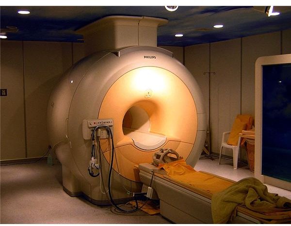
Above right: Modern MRI machine. (Supplied by Braegel at Wikimedia Commons; GNU Free Documentation License; https://upload.wikimedia.org/wikipedia/commons/b/bd/Modern _3T_MRI.JPG)
The Reason MRI Technology Works
The fundamental mechanisms and workings of MRI machines rely on principles of quantum mechanics. Each subatomic particle, such as a proton, has a certain level of spin. Exactly as the name suggests, these particles spin at a certain rate, either up or down. By adjusting the magnetic field surrounding these subatomic particles, the spin rate is altered. As the spin returns to normal, the MRI machine is able to determine its location . By locating a plethora of these subatomic particles, an image of tissues can be developed, giving researchers a picture of the subject’s particular area.
Structure of the MRI Scanner
The primary component of an MRI machine is the scanner. This is a device, usually shaped like a C, which creates the magnetic field and identifies the location of the subatomic particles. A magnet is the base component of the scanner, with additional parts built around the magnet. The majority of electromagnets used in MRI machines are made from niobium-titanium, an alloy that must be cooled by liquid helium kept at -492-degrees Fahrenheit (-269-degrees Celsius). The liquid helium is encased in a protective shield and then covered by a layer of thermal shielding to protect the subject from the extreme cold. Additional components are necessary to make sure the magnetic field is properly utilized. Shim and gradient coils are used to correct flaws in the scan and localize the magnetic resonance and radio frequently signals.
Dangers of MRI Machine Mechanisms
On occasion, deaths and injuries have resulted from the mechanisms and workings of MRI machines. Due to the fact that a strong magnetic field is created within the machine, any metallic device that is magnetic can be turned into a projectile within the scanning tube. Also, any electromechanical devices such as pacemaker can be directly damaged by the magnetic field and radio frequency system. In order to prevent these sort of problems caused by the mechanisms and workings of MRI machines, physicians should conduct a concrete investigation into a patient’s medical history before performing an MRI scan.
Resources
“Advances in MRI Equipment Design, Software, and Imaging Procedures” AAPM: https://www.aapm.org/meetings/amos2/pdf/26-5961-46702-744.pdf
“MRI” Mayo Clini: https://www.mayoclinic.com/health/mri/MY00227


