Ultrasound is one of the oldest forms of medical imaging, It can be used to uncover internal anatomy and to study blood flow throughout the body. Though best known for its use in pregnancy, it has many other applications and is an area of emerging technology.
How ultrasound imaging works
Ever since Dr. George Ludwig used naval sonar technology to detect gallstones in the late 1940s, medical ultrasound imaging has been a major tool in the diagnostician’s box. It is one of the oldest medical imaging modalities, predated only by X-ray imaging, but ultrasound technology continues to improve, allowing amazing real-time studies.
An ultrasound image starts with a hand-held device called a transducer, which is held against body tissue such as the skin. The transducer produces sound at frequencies far beyond the range of human hearing. To image more superficial tissues, higher frequencies (usually from about 7 to 18 MHz) are used to allow the greatest possible resolution; for deeper tissues, a lower frequency (usually from 2 to 6 MHz) is used, which trades off resolution for deeper penetration. A water-based gel to allow the transmission of sound from the transducer into the body tissues.
The transducer detects the echoes of the sounds it produces. The time it takes the echoes to return as well as the strength of the echoes provide information that is converted into an image. Because it provides good-quality images of soft tissues, sonography is used for clinical diagnosis in many fields of medicine, including obstetrics, cardiology, gastroenterology, and opthalmalogy.
Images:
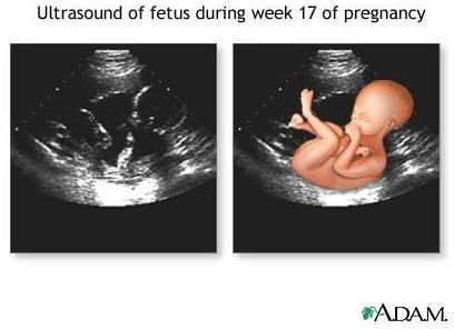
Left: Sonogram of a 17-week fetus. The drawing superimposed on the right shows the fetal outline. Source: NIH.gov
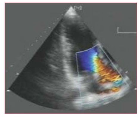
Center: Echocardiogram showing color-flow mapping of blood flow. Source: NIH.gov
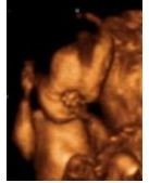
Right: 3D ultrasound of a 32-week fetus. Source: Adapted from en.Wikipedia.org, work by User:DrJoe K, licensed under Creative Commons Attribution 3.0
Sonogram images
Uses of ultrasounds during pregnancy
Sonography is used extensively, and most famously, to produce images of the embryo or fetus during pregnancy. Ultrasound uses no ionizing radiation and is considered completely safe for both mother and baby.
Physical measurements taken during first-trimester ultrasounds are more accurate than calculations based on last menstrual cycle for determining gestational age. These early sonograms are usually conducted transvaginally, while second- and third-trimester sonograms are conducted transabdominally. A thorough ultrasound of the fetus around 18-20 weeks gestation is a standard part of prenatal care in many places. This procedure can detect fetal abnormalities and determine the sex of the fetus. Ultrasounds may also be used in the later stages of pregnancy to diagnose placenta previa, determine fetal position, and obtain other clinically significant information on the pregnancy.
Doppler sonography and echocardiograms
Doppler sonography makes use of the Doppler effect, the observation that a wave source’s frequency is affected by its motion relative to the observer. Doppler sonography allows imaging of the direction, speed, and location of blood flow. Modern ultrasound technology mainly uses pulsed-wave (PW) Doppler. PW ultrasound machines transmit sound in pulses, rather than in a continuous wave. The time between the issuing of a pulse and the reception of its echo can be used to calculate the distance to the target by taking into account the speed of sound through body tissue. Software can detect Doppler shifts at the same time.
Doppler sonography is used in echocardiograms, also called cardiac ultrasounds. The echocardiogram is one of the most widely used diagnostic tests in cardiology, and is especially useful for diagnosing and assessing diseases of the heart valves. This test can assess the heart’s size and capacity, the existence and the severity of coronary artery disease, and any damage to heart muscle tissue. It is also used to evaluate surrounding structures such as the pericardium and aorta. The use of Doppler technology allows detection of problems that affect blood flow through the heart, such as backward blood flow through the valves (regurgitation) and septal defects (holes in the interior heart wall).
3D and 4D ultrasounds
Three-dimensional ultrasound technology involves sending sound waves in multiple directions into the body. Sophisticated medical imaging software then reconstructs the echoes into a three-dimensional volume representation. 3D sonograms are used both in obstetrics and in echocardiograms. A “four-dimensional ultrasound” is a marketing term for a 3D ultrasound conducted in real time (the fourth dimension) to create a moving image.
Careers in ultrasonography
Sonographers need not be medical doctors. They receive specialized medical imaging training in anatomy and in the use of ultrasound machines. Sonographers (also called ultrasonographers and ultrasound technicians) are skilled anatomists whose training allows them to use the ultrasound machine as an extension of their own senses to detect anatomical abnormalities. Each country has its own certification program for sonographers.


