The Gram stain is one of the most basic tests in diagnostic microbiology. It divides bacteria into two categories: Gram-positive and negative cell walls. This information is highly significant in describing bacterial species.
Characteristics
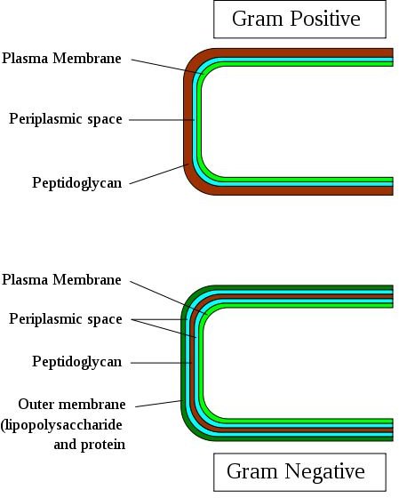
The distinguishing characteristic of Gram positive bacterium is the composition of its cell wall – several peptidoglycan layers joined together forming a thick and rigid structure. By contrast, gram-negative bacteria have only a thin peptidoglycan layer.
Moreover, the cell wall of a Gram-positive bacterium has teichoic acids, which mainly consist of alcohols (like ribitol and alcohol) and phosphate. Teichoic acids have two types: lipoteichoic acid and wall teichoic acid. Lipoteichoic acid traverses the peptidoglycan layer and is physically connected to the plasma membrane; wall teichoic acid does not traverse the peptidoglycan layer and is physically connected to this layer alone, never touching the plasma membrane. Both types of teichoic acids are negatively charged because they contain the phosphate group in their molecular structure. Because of the nature of their charges, they play a role in binding and regulating the movement of positive ions (cations) into and out of the cell. They also help in promoting cell growth as well as preventing extensive wall damage and possible cell lysis. Lastly, teichoic acids contribute to the bacterial cell wall’s antigenic specificity, making it very possible to identify gram positive bacteria in various laboratory tests. (Shagam 2006; Wheelis 2007)
Grouping medically important types of gram-positive streptococci is now a possibility because different strains of the bacteria are covered with different polysaccharides. The differences in polysaccharides that compose their cell walls are therefore a very important factor in accurately identifying streptococcus species, especially the pathogenic ones. Every streptococcus species is unique because the types and structural configuration of its cell wall polysaccharides may be different. A bacterium with a cell wall composed of only 40% peptidoglycan can be considered gram positive so long as it can be stained with the gram stain. An example is the Mycobacterium which has a cell wall consisting of 60% mycolic acid (a waxy lipid), with the rest (40%) being peptidoglycan. (Shagam 2006; Wheelis 2007)
Characteristics of Gram-Negative Bacterial Cell Walls
The presence of an outer membrane and the possession of only few peptidoglycan layers in the cell wall distinguish Gram-negative bacteria from Gram-positive ones. Lipids covalently linked to proteins called lipoproteins are the molecules that bind the peptidoglycan to the outer membrane. The peptidoglycan is located in the periplasm, a space filled with fluid located between the plasma membrane and the outer membrane. A high amount of degradative enzymes and transport proteins are found in the periplasm. Unlike Gram-positive cell walls, we cannot find teichoic acids in the Gram-negative cell walls. In addition, the cell walls of Gram-negative bacteria are more prone to mechanical breakage because of the low amount of peptidoglycan. (Shagam 2006; Wheelis 2007)
The outer membrane of a Gram-negative bacterium is composed of lipopolysaccharides (LPS), phospholipids, and lipoproteins. It plays a very important role in the survival of the bacterium under environmental pressure. For example, it prevents the whole bacterium from being phagocytosed (e.g. by macrophages) because of its strong negative charge. The mechanism of evading phagocytosis involves a complex biochemical pathway which is not the scope of this article. Furthermore, the outer membrane serves as a barrier for the bacterium against the destructive effects of various antibiotics (e.g. erythromycin, penicillin, amoxicillin), digestive enzymes like lysosomal enzymes, heavy metals, detergent substances, bile salts, and several dyes. (Shagam 2006; Wheelis 2007)
It is, however, necessary that the outer membrane is not totally restrictive to the entrance of molecules because it may jeopardize the health of the bacterium if important nutrients could not enter the cytoplasm. The outer membrane is actually permeable to nutrients due to the presence of porins, proteins that form channels toward the cytoplasm. Porins allow the entry of valuable molecules like disaccharides, nucleotides, peptides, amino acids, iron, and vitamin B12 but it prevents the entry of other molecules, especially the bigger ones.
The polysaccharide components of outer membrane’s LPS serves as bacterial antigens and are very helpful in identifying species of Gram-negative bacteria in the laboratory. There are certain laboratory tests that detect antigens specific for a single species. The LPS is therefore significant in medical diagnosis of pathogenic infections. (Shagam 2006; Wheelis 2007)
Graphical illustration of the 2 types of bacterial cell walls
References
- Wheelis, Mark. 2007. Principles of Microbiology. Jones & Bartlett Publishers.
- Shagam, Janet. 2006. Introduction to Microbiology. Pearson Education Limited.
Photo credit: https://en.wikipedia.org/wiki/File:Gram-Cell-wall.svg


