Agenesis of the corpus callosum occurs when the nerves connecting the two brain hemispheres fail to form properly. Consequences of this malformation range from neurologically normal individuals to mental retardation.
What is the corpus callosum?
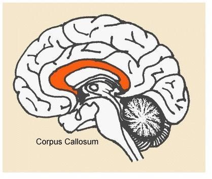
This neuroanatomical landmark (pictured below in orange) is located in the center of the brain. It develops from the 5th to the 17th weeks of pregnancy. Lying within the longitudinal fissure, it is a bundle of nerve fibers that runs between and connecst the two cerebral hemispheres. It is through this nerve bundle that the two halves of the brain communicate. Both myelinated and nonmyelinated nerve axons comprise this structure.
The Corpus Callosum
Failure to Form Properly
Agenesis of the corpus callosum (ACC) is a malformation of the brain that occurs in the developing fetus. Its cause is unknown, and most cases are sporadic (that is, not familial). There are rare instances of inheritable autosomal dominant, autosomal recessive, or x-linked recessive genetic mutations that cause this malformation. ACC has also been seen in some cases of specific metabolic conditions, including pyruvate dehydrogenase deficiency and nonketotic hyperglycinemia. This birth defect may be associated with other conditions, including Arnold-Chiari malformation, Dandy-Walker syndrome, Andermann syndrome, schizencephaly, holoprosencephaly, and Aicardi’s syndrome. Children with ACC may also have other malformations, such as facial or cardiac birth defects. The incidence of ACC is 0.03% to 0.7% of the general population, and it accounts for 2-3% of mentally disabled persons.
Neurological Consequences of ACC
There is a wide variety of signs and symptoms associated with this condition, and the presentation differs based on the extent to which this neural structure forms. Common consequences include seizures, delayed development, microcephaly (small brain size), and mental retardation. However, evidence of this malformation has also been found in the brains of seemingy normal people. Thus, its effects are somewhat unpredictable. People with ACC may have normal intelligence, but further testing could reveal mild abnormalities in higher cognitive function, such as pattern matching.
Diagnosis, Prognosis, and Treatment
Diagnosis of ACC is done through imaging of the brain, which reveals a complete or partial abscence of these nerve fibers in the center of the brain. A prenatal sonogram can allow for early diagnosis and prenatal counseling during pregnancy. Prognosis and treatment depend on the severity of neurological effects. Treatment is generally symptom-based, and there is no curative procedure.
References
Moe Paul G, Benke Timothy A, Bernard Timothy J, Levisohn Paul, “Chapter 23. Neurologic & Muscular Disorders” (Chapter). Hay WW, Jr., Levin MJ, Sondheimer JM, Deterding RR: CURRENT Diagnosis & Treatment: Pediatrics, 19e: https://www.accessmedicine.com.ezproxy.galter.northwestern.edu/content.aspx?aID=3405101 .
“NINDS Agenesis of the Corpus Callosum Information Page.” National Institute of Neurological Disorders and Stroke, National Institutes of Health, Bethesda, MD. https://www.ninds.nih.gov/disorders/agenesis/agenesis.htm
Penny SM. Agenesis of the corpus callosum: neonatal sonographic detection. Radiol Technol. 2006 Sep-Oct;78(1):14-8. Review.
Waxman SG, “Chapter 10. Cerebral Hemispheres/Telencephalon” (Chapter). Waxman SG: Clinical Neuroanatomy, 26e: https://www.accessmedicine.com.ezproxy.galter.northwestern.edu/content.aspx?aID=5273317 .
Image Credit
Jones, Paul. The Multiple Sclerosis Information Trust: “All About Multiple Sclerosis” https://www.mult-sclerosis.org/corpuscallosum.gif


