Learn About Zygomatic Bone and Zygomatic Arch
Description
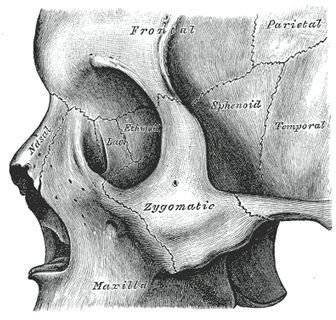
The zygomatic bone, also called the malar bone, is a roughly trapezoid-shaped facial bone located under each eye, forming the lower outside edge of the orbit (eye socket). A process (extension or outgrowth) of the zygomatic bone meets with a process of the temporal bone (located at the side of the skull at the temple), forming the horizontal zygomatic arch. Both the zygomatic bone and the zygomatic arch may be called the zygoma. They are colloquially known as the “cheekbone,” which is prominently visible on the faces of some people. At right: Cheekbone anatomy from Grey’s Anatomy.
Function
Besides supporting the side wall and floor of the orbit, the zygomatic bone also articulates with (i.e. connects to) a number of other facial bones. These include the maxilla (the upper jawbone that contains the upper teeth), the frontal bone (the “forehead bone” that forms part of the braincase and orbit), the sphenoid bone (located “under” or behind the zygomatic arch), and of course, the temporal bone, with which it forms the zygomatic arch. The zygomatic arch supports the main jaw muscle, the masseter, which is necessary both for biting and chewing food and for speech. Several other facial muscles attach to the zygomatic bone, as well.
Medical Considerations
In facial fractures, the fracture line commonly passes through the junction between the zygomatic and sphenoid bones and continue to the junction between the zygomatic and frontal bones. These fractures are called zygomatic complex fractures and may be complex to repair surgically because of the potential for damage to the eye. People of Asian descent typically have a prominent zygoma. Occasionally, Asian people may request elective cosmetic surgery to reduce the size of the cheekbone.
Evolution of the Zygomatic Arch
In comparative anatomy, the zygomatic bone is also called the jugal bone, which is found in all tetrapods (amphibians, reptiles, birds, and mammals). In the ancestors of mammals, the synapsid reptiles, a single opening was present in the skull behind the eye socket. The jugal (zygomatic) bone stretched from the bottom of the eye socket to the bottom of this opening. Around the time the first mammals evolved, the vertical separation between this opening and the orbit disappeared, leaving the zygomatic arch formed from the zygomatic bone and temporal bone. A vertical connection between the zygomatic bone and frontal bone is again present in humans, but the zygomatic arch remains.
References
- Adam J. Cohen et al. “Facial Trauma, Zygomatic Complex Fractures.” 2008, Medscape Online Medical Encyclopedia.
- Babak Jahan-Parwar and Keith Blackwell. “Facial Bone Anatomy.” 2009, Medscape Online Medical Encyclopedia.
- Kenneth V. Kardong. Vertebrates: Comparative Anatomy, Function, Evolution. 1995; Dubuque:Wm. C Brown Publishers.
- T. Wang et al. “Reduction Malarplasty with a New L-Shaped Osteotomy through an Intraoral Approach: Retrospective Study of 418 Cases.” Plast Reconstr Surg. 2009 Oct;124(4):1245-53.