History of the CT Scanner - X-Rays, Contrast Agents and Radiation Risks with the Axial Scanning Machine
What is Computed Tomography?
Within the medical industry, computed tomography (CT) is used to image sections of a person with the use of energy waves. The basic way a CT scanner works is by taking a series of two-dimensional X-rays of a patient in a circular format. These X-rays are then placed together through a digital geometry process to create a thee-dimensional image. This has the benefit of allowing physicians and medical practitioners to be able to look through the layers of images to find a possible problem in the patient. Essentially, organs and tissues can be represented on a computer screen as they look in the body. While this technique is most commonly used in the medical industry, throughout the history of the CT scanner, other fields of research have also implemented the technology.
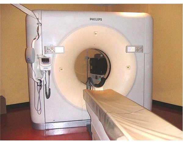
Above right: CT Scanner. (Supplied by Glitzy queen00 at Wikimedia Commons; Public Domain; https://upload.wikimedia.org/wikipedia/commons/2/23/64_slice_scanner.JPG)
Origins of the CT Scanner
While the concept of tomography in medicine was first invented by Italian radiologist Alessandro Vallebona in the early 1900s, the method at the time was simply to take a series of X-rays to attempt to create a slice of a patient’s body. This was used for decades until Sir Godfrey Hounsfield of EMI Central Research Laboratories in the United Kingdom used the concept in conjunction with computer technology and an axial scanning method. He built a commercially viable CT scanner in 1972 based on his research, which also implemented design elements from Allan McLeod Cormack of Tufts University in Massachusetts. Both Hounsfield and Cormack shared the 1979 Nobel Prize for Medicine due to the invention, changing the direction of internal diagnostics and beginning the long history of the CT scanner.
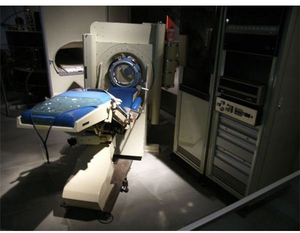
Above left: Early design for a CT scanner. (Supplied by Gdh at Wikimedia Commons; Public Domain; https://upload.wikimedia.org/wikipedia/commons/a/ae/Emi1010.jpg)
Advantages and Disadvantages of CT Scans
There are a number of advantages and disadvantages to the design of the device. Throughout the history of the CT scanner, new methods of advancing the technique have come at the cost of increased radiation exposure to the patient. A CT scanner used the concept of multiplanar reformatted imaging, a system that allows researchers to view the slice of biological material on a multitude of geometric planes. As the resolution is increased with each generation, higher doses of radiation are needed to to conduct the complex scans. While radiation is constantly being reduced in other medical testing equipment, CT scanning has increased the volume of radiation patients are exposed to in general within the industry.
Importance of Using CT Scanners
The history of the CT scanner as an important instrument of medical testing was greatly advanced due to the introduction of contrast agents. Essentially, a form of dye is intravenously administered into the patient to provide better image quality in the scan. While very few patients suffer allergic reactions, some situations have been known to cause severe problems. In addition, common side effects include nausea and warmth in the pelvic area. This is due to the fact that the contrast agent has detrimental effects on the kidneys of patients, sometimes leading to renal issues.
Resources
“Brief History of CT Scanners” Imaginis: https://www.imaginis.com/ct-scan/history.asp
“CT Scan” eMedicineHealth: https://www.emedicinehealth.com/ct_scan/article_em.htm