How Does the Circulatory System Work - An Overview
What is the Circulatory System?
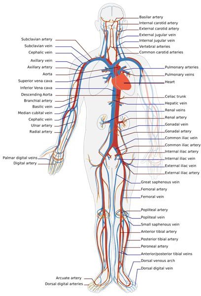
The circulatory system is an organ system of the human body. It is primarily responsible for transporting water, nutrients and waste, including oxygen and carbon dioxide, throughout the body. This is done by a complex system of blood vessels which include arteries, veins and capillaries. Arteries and capillaries are responsible for carrying blood away from the heart to the rest of the body and its organs, whereas veins are responsible for carrying blood from the rest of the body and its organs to the heart. The blood carried by the arteries is oxygenated while blood carried by the veins is deoxygenated. Once the veins carry blood to the heart, which is at the center of the circulatory system, the blood gets reoxygenated for transport back to the body via the arteries. Exchange of gases, nutrients and wastes occurs through the walls of the capillaries between the blood and the various tissues and organs. The capillaries are part of the microcirculation network within the body. The overall maintenance of homeostasis is also achieved by the circulatory system through a number of control mechanisms. We will look at how does the circulatory system work.
Different Types of Circulatory Systems
A large number of invertebrates do not possess a circulatory system. The cells within their bodies are close to the environment for gases, nutrients and wastes to move in and out of the cells by diffusion. Land animals have multiple layers of cells and possess a different circulatory system from invertebrates. Essentially, there are two types of circulatory systems in higher animals. They are the open circulatory system and the closed circulatory system. We will look at each one briefly.
-
Open Circulatory System: The heart and capillaries are lacking in this type of system. Blood vessels, which are joined to open sinuses, exist that push the blood along like pumps. Blood is forced from the vessels into the sinuses, where it bathes the organs. At the same time, various other vessels receive blood from the sinuses and send it back again to the vessels for pumping. This type of system is found in arthropods such as insects and spiders, as well as some mollusks. The system is not very efficient and there are many limitations to it when compared to the closed circulatory system.
-
Closed Circulatory System: In this type of system, blood flows through a closed system of blood vessels. The blood vessels include arteries, veins and capillaries. Diffusion takes place between the capillaries and the tissues and organs of the animal of the various nutrients, gases and wastes. Closed circulatory systems in humans have a functioning heart that acts as the primary pump for the blood. There are different types of closed circulatory systems found even in vertebrates. For example, fishes possess a system where the heart is two-chambered, frogs have a closed circulatory system with a three-chambered heart and humans have a four-chambered heart.
The circulatory system in humans has three main components to it. These are the pulmonary circulation, systemic circulation and coronary circulation. We will first discuss the anatomy and functioning of the heart, and then we will examine the various aspects of the circulatory system.
Heart Anatomy and Function
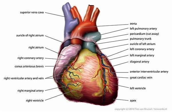
The heart is a muscular organ within the body that pumps blood from the veins into arteries in order to ensure proper flow of blood throughout the circulatory system. The wall surrounding the heart has three layers. The outer layer is the epicardium, the middle layer is the myocardium and the inner most layer is the endocardium. The epicardium is mostly comprised of connective tissue and mainly functions as a protective layer around the heart. It is considered part of the pericardium. The myocardium, or middle layer of the heart, consists mostly of cardiac muscle. Cardiac muscle is responsible for propelling blood out of the chambers of the hearts into the pulmonary circulatory system and the systemic circulatory system. The endocardium is the inner most layer of the heart and is made up of epithelial tissue. The epithelial tissue forms a lining of the inner surface of the heart chambers and valves. The endocardium is primarily responsible for myocardium function.
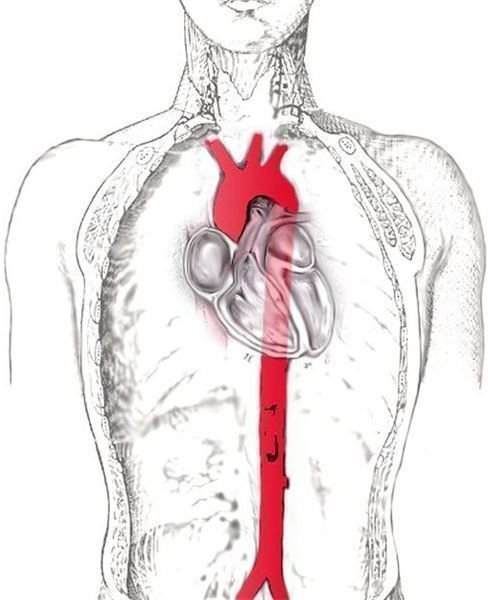
The human heart is a four-chambered organ. The heart is divided into two cavities by a wall of muscle known as the septum. Blood is pumped by the left cavity to the body, while the right cavity pumps blood through the lungs. The chambers making up the cavities are known as atria, which are located on top, and the ventricles which are at the bottom. The heart is also composed of various arteries and veins that carry blood to and from the heart. The pulmonary arteries carry blood to the lungs while the pulmonary veins are responsible for bringing blood from the lungs back to the heart. The aorta is the largest artery in the body and originates in the left ventricle. It carries oxygenated blood throughout the body. The aortic arch of the heart contains the carotid arteries, the subclavian arteries, the brachiocephalic artery, the coronary arteries, the iliac arteries and the renal arteries. Carotid arteries supply the head and neck region of the body with oxygenated blood, the subclavian arteries supply blood to the left and right arms of the body, the brachiocephalic artery supplies blood to the right arm and the head and neck, and the coronary arteries supply blood to the myocardium. The iliac arteries branch out in the lower trunk to supply blood to the thighs and legs. Blood to the kidneys is supplied by the renal arteries whereas the celiac axis and the superior and inferior mesenteric arteries supply blood to the liver, spleen and intestines.
The superior vena cava is the vein from which the blood enters the heart from the head and the arms. The inferior vena cava is where the blood comes to the heart from the body and legs. The mitral valve and the tricuspid valve, collectively known as the atrioventricular valves, lie between the atria and the ventricles and control the flow of blood into the heart. The aortic valve and the pulmonary valve are collectively known as the semilunar valves. These valves allow blood to enter the arteries, but prevent backflow of blood into the ventricles from the arteries.
The Pacemaker of the Heart
A pacemaker is a group of excitable cells whose firing is rhythmic. The pacemaker portion of the heart is responsible for electrical activity. The sinoatrial node (SA) is the pacemaker of the heart. Special muscles within the sinoatrial node are responsible for contraction. The contraction of the heart muscle is produced due to the action potential within the cells.
Pulmonary Circulation
Pulmonary circulation refers to the movement of blood from the heart to the lungs and back to the heart again. Blood that is deoxygenated leaves the heart and travels to the lungs. Blood that is oxygen-depleted leaves the systemic circulation and enters the right atrium through the superior vena cava and the inferior vena cava. The tricuspid valve pumps blood into the right ventricle followed by the semilunar valve that pumps blood into the pulmonary artery. The pulmonary arteries carry the deoxygenated blood to the lungs. Once the blood reaches the lungs it releases carbon dioxide and picks up fresh oxygen. The blood leaves the lungs via the pulmonary veins and travels back to the heart and enters the left atrium. This is the end of the pulmonary cycle. From here, the freshly oxygenated blood is pumped by the left atrium through the bicuspid valve or mitral valve into the left ventricle. Distribution of the blood throughout the body takes place through systemic circulation before returning to the pulmonary circulation system.
Systemic Circulation
Systemic circulation is where blood leaves the heart and enters the cells of the body and then returns back to the heart. The lungs are the only organs that are not part of the systemic circulation system. Oxygenated blood enters the systemic circulation through the left ventricle. As the ventricle contracts, blood is forced into the aortic arch and then into the aorta. Once in the aorta, blood travels to the cardiac muscle through the coronary arteries, to the head and neck through the carotid arteries, to the arms through the subclavian arteries and the brachiocephalic arteries, to the kidneys through the renal arteries and to the thighs and legs via the iliac arteries which branch out to form the femoral and popliteal arteries supplying the lower trunk of the body. The liver, spleen and intestines are also supplied with blood through major arteries from the heart. Capillaries form venules, which in turn form veins. The inferior vena cava along with the superior vena cava return blood that is low in oxygen to the right atrium of the heart.
Coronary Circulation
Coronary circulation primarily involves circulating blood through a network of blood vessels to the myocardium. Deoxygenated blood is also removed from the heart by the cardiac veins. The coronary arteries are mainly responsible for supplying blood to the heart muscle. The left and right coronary arteries are the main arteries of the coronary circulation system. The left coronary artery branches out into the left anterior descending artery (LAD) and the left circumflex artery (LCX). The LAD supplies blood to the anterolateral myocardium, apex and the interventricular septum. The LCX supplies blood to the posterolateral ventricle and the anterolateral papillary muscle. The right coronary artery branches out into the right marginal artery and the posterior descending artery. The right coronary artery supplies blood to the right ventricle and also about 25-35% to the left ventricle. The cardiac veins are responsible for carrying deoxygenated blood to the right atrium.
Heart Diseases
Heart diseases are a major health concern around the world. Different types of heart diseases exist. Thickening and hardening of the artery walls as a result of deposition of fatty materials is known as arteriosclerosis. The same process affects the aorta and other major vessels and is known as atherosclerosis. Many illnesses that are indirectly related to the heart can also occur if the circulatory system is not functioning well. These include high blood pressure, aneurysms and stroke.
Steps for Prevention
In order to prevent heart diseases and to maintain a properly functioning circulatory system, certain steps can be taken. One is to avoid smoking any form of recreational drugs, especially tobacco. Another is to eat healthy and try and minimize on greasy and oily foods. Drinking sufficient amounts of water is also important. One should also exercise regularly to keep the circulatory system functioning efficiently. Going for regular walks, doing aerobics or jogging are all good forms of cardiovascular exercises. Going for routine checkups at the doctor’s office is also a good way to ensure that one is healthy and that the heart and in turn the circulatory system are in good working condition.
References
1. Natural Health School: https://www.naturalhealthschool.com/8_1.html
2. The Internet Encyclopedia of Science: https://www.daviddarling.info/encyclopedia/C/circulatory_system.html
3. Access Excellence: https://www.accessexcellence.org/AE/AEC/CC/heart_background.php
Image Credits:Wikimedia Commons/JHeuser/Tvanbr/LadyofHats